12 February 2020
A novel technique that isolates individual gut cells from mice with intestinal parasites may yield new insights on the immune response to these infections.

A new technique may help scientists study the body’s immune response to intestinal parasite infections one gut cell at a time, according to a study published today in eLife.
Intestinal parasites are a serious threat to both humans and livestock in large parts of Africa, South America and Asia. The findings may help scientists understand how adult parasites evade the immune system and to test ways to boost the immune response to fight these infections.
A parasite’s larvae may infect humans and other organisms through contaminated food or by penetrating bare skin. Once the parasites grow into adults inside the body, the immune system has a hard time getting rid of them and scientists have found it challenging to study immune cells in infected tissues.
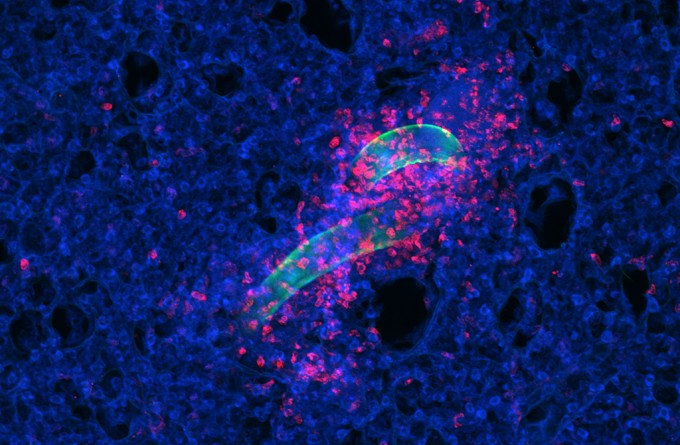
“One big problem has been the difficulty to extract immune cells from an infected gut, as the infection causes very strong local reactions such as intense cellular slime production to try and flush the worms out,” says senior author Johannes Mayer, PhD, Research Fellow at the Malaghan Institute of Medical Research.
Mayer and colleagues tested many different ways to extract immune cells from the guts of mice infected with an intestinal parasite called Heligmosomoides polygyrus bakeri. Most of their attempts failed, but they eventually developed a technique that isolated millions of immune cells from the infected animals’ guts. The technique involves three washes with EDTA, an agent to remove the mucus, lasting for 10 minutes. This is followed by 30 minutes in a solution of enzymes that help break down the tissue into individual cells, and then cell filtration.
“This allowed us to study the individual immune cells for the first time,” explains lead author Laura Ferrer-Font, PhD, Staff Scientist at the Malaghan Institute. “We used a new technology called spectral flow cytometry to look at many different types of immune cells all at the same time, and identified various changes that take place throughout the course of parasitic infection.”
The team also verified that the immune changes they saw in the cells were representative of the immune changes that occurred in the tissue taken from the infected animals, ensuring that the cell-extraction process did not skew their findings.
Read this article in our quarterly newsletter Scope 71: COVID-19: Moving New Zealand towards a cure
“Now that we have found a way to extract immune cells from parasite-infected guts, we can start to answer important questions about the immune response,” Mayer concludes. “This technique will enable scientists to use powerful tools like single-cell RNA sequencing to study the immune response in different hosts. It might also help those studying inflammatory bowel disorders or food allergies to extract single cells from the gut for further investigation.”
The paper ‘High-dimensional analysis of intestinal immune cells during helminth infection’ can be freely accessed online at https://doi.org/10.7554/eLife.51678. Contents, including text, figures and data, are free to reuse under a CC BY 4.0 license.
This article is a media release provided to us by eLife.
Related articles

In Focus: Mapping the lung's fight – how the entire organ responds to infection
18 April 2024

New research suggests hookworms could offer protection from severe Covid symptoms
14 August 2023

In Focus: From tiger conservation to parasites and the allergy epidemic
10 May 2023

In focus: Making friends with enemies, unexpected solutions to our allergy epidemic
16 February 2022
Could a dose of worms be the answer to treating incurable autoimmune diseases?
14 November 2021

Study show gut worms protect against development of allergic conditions in the skin
7 July 2020