
The Ronchese Laboratory is investigating the fundamental biology behind the development of allergies and inflammatory conditions. By better understanding the genetic and environmental triggers that are responsible for the development of allergies, they aim to find opportunities prevent these diseases from developing in the first place.
The Lab's current focus is on dendritic cells, a rare type of immune cells whose function is to initiate various immune responses. Dendritic cells are found in most tissues in our bodies and especially in the skin, the intestinal tract, and the airway. The team has perfected techniques to label allergens with fluorescent markers, monitor their uptake by dendritic cells in vivo, and follow their transport to lymph nodes where they initiate immune responses. By using these fluorescence-labelled antigens in combination with flow cytometry and transcriptomics, the Ronchese Lab is able to characterise in detail how allergen uptake alters the properties of dendritic cells and endows them with the ability to initiate Th2 immune responses. Results from these studies show that allergens impact the immune system at several levels, acting on dendritic cells directly, and also indirectly via the induction of innate cytokines in the tissue environment. By carrying out functional experiments, the group can alter or block the signalling of these innate cytokines in dendritic cells, assess their contribution to dendritic cell development, their expression of key mediators, and their priming of Th2 immune responses and subsequent allergic inflammation.
To increase the specificity of their models, the Ronchese Lab is also comparing allergen-treated dendritic cells to dendritic cells that have been exposed to other micro-organisms, such as bacteria and fungi, that prime Th responses of different phenotypes such as Th1 or Th17. They plan to compare the transcriptomes of dendritic cells exposed to these various stimuli to those of dendritic cells exposed to allergens. They expect that these studies will enable them to define the dendritic cell “Th2 signature” with a high level of precision.

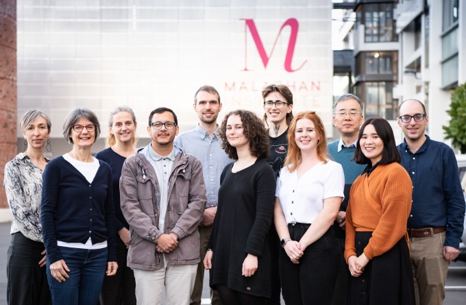
Dr Olivier Lamiable
Team Leader

Dr Kerry Hilligan
Team Leader

Dr Sou Ochiai
Postdoctoral Research Fellow
Senior Research Officers: Marie Sophie-Fabre, Evelyn Hyde, Shiau-Choot Tang
Research Officers: Abbie Larson, Rebecca Palmer, Sam Old
PhD Student: Greta Webb
Research areas
- Allergic and inflammatory disease
- Immune health
- Transcriptomic analysis of immune cells
Research projects
- Functional and transcriptomic plasticity of dendritic cells during allergic and non-allergic immune responses
- Role and regulation of the CCL17-CCL22-CCR4 axis in allergic priming
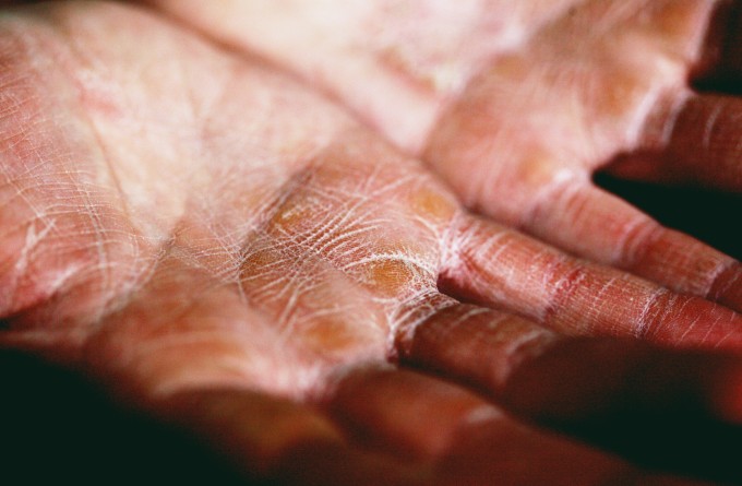
- The skin as an important site of allergic sensitisation
Collaborations
Featured articles
Discovery points to the skin as ‘ground zero’ for allergic disease
19 November 2021
Royal Society Te Apārangi Fellowship to investigate relationship between germs and the development of allergies
2 November 2021
HRC grant to investigate the link between dendritic cells and allergic skin disease
27 July 2021
Allergy research honours in Burnet Oration
29 April 2019
Featured publications
Mayer JU, Hilligan KL, Chandler JS, Eccles DA, Old SI, Domingues RG, Yang J, Webb GR, Munoz-Erazo L, Hyde EJ, Wakelin KA, Tang SC, Chappell SC, von Daake S, Brombacher F, Mackay CR, Sher A, Tussiwand R, Connor LM, Gallego-Ortega D, Jankovic D, Le Gros G, Hepworth MR, Lamiable O, Ronchese F (2021). Homeostatic IL-13 in healthy skin directs dendritic cell differentiation to promote TH2 and inhibit TH17 cell polarization. Nat Immunol. 22(12):1538-1550.
Hilligan KL, Tang SC, Hyde EJ, Roussel E, Mayer JU, Yang J, Wakelin KA, Schmidt AJ, Connor LM, Sher A, MacDonald AS, Ronchese F (2020). Dermal IRF4+ dendritic cells and monocytes license CD4+ T helper cells to distinct cytokine profiles. Nat Commun. 11(1):5637.
Blecher-Gonen R, Bost P, Hilligan KL, David E, Salame TM, Roussel E, Connor LM, Mayer JU, Bahar Halpern K, Tóth B, Itzkovitz S, Schwikowski B, Ronchese F, Amit I (2019) Single-Cell Analysis of Diverse Pathogen Responses Defines a Molecular Roadmap for Generating Antigen-Specific Immunity. Cell Systems 8(2):109-121.e6.
Ochiai S, Jagot F, Kyle RL, Hyde E, White RF, Prout M, Schmidt AJ, Yamane H, Lamiable O, Le Gros G, Ronchese F. (2018) Thymic stromal lymphopoietin drives the development of IL-13+ Th2 cells. Proc Natl Acad Sci U S A. 115(5):1033-1038.
Connor LM, Tang SC, Cognard E, Ochiai S, Hilligan KL, Old SI, Pellefigues C, White RF, Patel D, Smith AA, Eccles DA, Lamiable O, McConnell MJ, Ronchese F (2017) Th2 responses are primed by skin dendritic cells with distinct transcriptional profiles. J Exp Med. 214(1):125-142.
Connor LM, Tang SC, Camberis M, Le Gros G, Ronchese F (2014) Helminth–conditioned dendritic cells prime CD4+ T cells to IL-4 production in vivo. J. Immunol. 193(6):2709-17.
Ochiai S, Roediger B, Abtin A, Shklovskaya E,Fazekas de St Groth B, Yamane H, Weninger‡ W, Le Gros G, Ronchese.F. (2014). CD326loCD103loCD11blo dermal dendritic cells are activated by Thymic Stromal Lymphopoietin during contact sensitization in mice. J Immunol. 193(5):2504-11.