2 May 2020
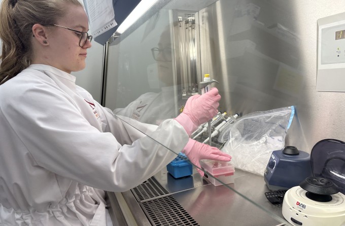
A new confocal microscope is a welcome addition to the Institute’s Hugh Green Cytometry Centre, offering an exponential shift in the depth and quality of images produced by experiments.

The more information gained from an experiment, the more scientific value it has. Any advancement in technology helps create better, more sophisticated data per experiment. It’s all part of creating a complete picture of what’s going on within a body in order to find ways intervene in the context of disease.
In line with this, the new confocal is a significant leap in the quality of information gained in a single microscopic image. Unlike regular microscopes which examine only a single layer of tissues or cells, the confocal creates multiple layers of images at different depths, effectively creating three-dimensional images. To this end, the new confocal has a super resolution module incorporated, which means we will be able to resolve structures down to 120nm in size, which is about 16 times smaller than bacteria. These features combined, along with a vastly improved processing speed, exponentially expand our understanding of the immune system in the context of the tissue sections.
“The new confocal has necessitated a change in the way our scientists think about their experiments,” says Kylie Price, Hugh Green Cytometry Fellow and Head of Research Technology. “Now we have the tools to ask really big picture questions and take a more holistic approach to big-data analysis.”
Read this article in our quarterly newsletter Scope 71: COVID-19: Moving New Zealand towards a cure
Related articles

In Focus: Shooting for the stars, propelling our research in the information age
21 March 2024
In Focus: Tailoring mRNA vaccines for immunocompromised populations
14 December 2023
Philanthropic partnership sees record $15 million boost to biomedical research
12 December 2023
Malaghan Institute and BioOra deliver automated manufacturing to scale up CAR T-cell cancer therapy in NZ
16 August 2023
In Focus: Using state-of-the-art technology to probe the immune system’s role in health and disease.
9 August 2023
Malaghan microscopy expert awarded advanced diploma
2 March 2023